
Our lab bridges endocrinology and nephrology to solve the complex challenges of kidney and cardiovascular health for youth and adults living with diabetes.
Research Laboratories
Leading-Edge Research Laboratories at UW Diabetes Institute: Advancing Understanding, Treatment, and Prevention of Diabetes and Related Diseases Through Innovation and Collaboration


Bjornstad-Pyle-Tommerdahl Laboratory

Bornfeldt Laboratory
The Bornfeldt laboratory is dedicated to understanding the cellular and molecular mechanisms of diabetes-accelerated cardiovascular disease, so that these complications can be effectively treated or prevented.
Bowen Laboratory
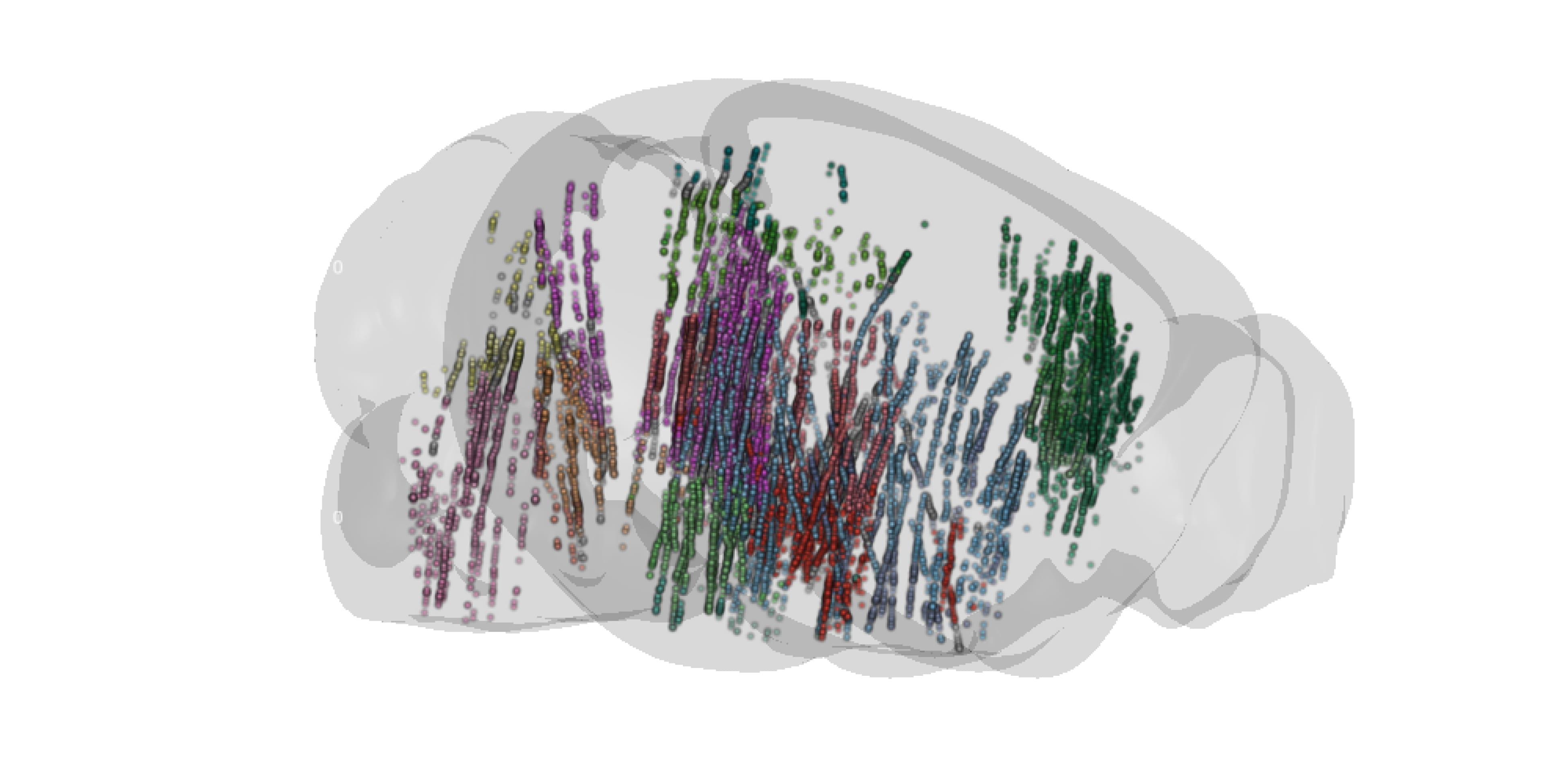
We study how brain-based predictions support homeostasis and prevent disease.

Capozzi Laboratory
The Capozzi laboratory is seeking to understand how hormones control nutrient metabolism in health and metabolic disease.
Cirulli Laboratory
The Cirulli laboratory is committed to understanding mechanisms of cell-cell and cell-matrix interactions in pancreatic islet cell development and function.

Crisa Laboratory
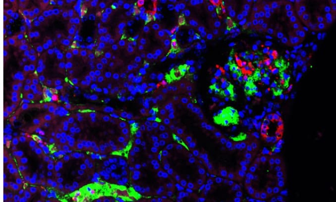
The Crisa laboratory seeks to understand the basic mechanisms by which blood vessels and myeloid immune cells may drive growth and regeneration of pancreatic islets during development and in diabetes.

Den Hartigh Laboratory
Dedicated to understanding the nutritional and hormonal impact of obesity and weight loss on adipocyte biology.

Dorfman Laboratory
The Dorfman laboratory investigates the protective role of sex steroids in hypothalamic inflammation and obesity.

Heinecke Laboratory
The Heinecke is dedicated to understanding the structure and function of HDL (good cholesterol), and to predicting the risk of cardiovascular disease in diabetic patients and other subjects at high risk of atherosclerosis.
Kanter Laboratory
The Kanter laboratory is focused on understanding the molecular mechanisms that result in complications of diabetes.

Kim Laboratory
The Kim laboratory focuses on nitric oxide (NO), a crucial molecule involved in maintaining vascular homeostasis.

Morton Laboratory
The Morton laboratory investigates how the brain regulates energy balance and glucose metabolism, and how disruptions in these processes contribute to obesity and diabetes.

Page Laboratory
The Page laboratory is dedicated to developing the first hormonal form of contraception for men.

Scarlett Laboratory
The Scarlett laboratory investigates the mechanisms whereby FGF1 action in the brain induces sustained remission of diabetic hyperglycemia.

Schur Laboratory
BBARC is dedicated to understanding brain regulation of appetite and obesity pathogenesis in humans

Schwartz Laboratory
The Schwartz laboratory investigates the role of the brain in the control of both energy balance and blood glucose levels with a focus on how defects in these systems contribute to the pathogenesis of obesity and diabetes.
Sewaybricker Laboratory
BBARC is dedicated to understand the brain regulation of appetite and body weight in children

Shao Laboratory
The Shao laboratory is dedicated to understanding the pathways for the generation of dysfunctional HDL and the roles of dysfunctional HDL in the pathogenesis of atherosclerosis, diabetes, and other inflammatory diseases in humans.

Sweet Laboratory
The Sweet laboratory investigates regulation and impairment of insulin secretion in type 2 diabetes; pancreatic beta cell death; metabolic basis of inflammation and T cell calcium metabolism in type 1 diabetes.
Thaler Laboratory
The Thaler laboratory seeks to discover new brain-based treatments for obesity and diabetes.

Vaisar Laboratory
The focus of the lab is on understanding molecular mechanisms by which lipoproteins contribute to development and progression of heart disease, stroke and Alzheimer’s disease. To facilitate these studies, we apply state-of-the-art quantitative mass spectrometry techniques to blood, CSF, cells and tissues.

Valencia Laboratory
We are dedicated to understanding the relationship between mitochondria and nutritional stressors in the context of metabolic disease. Within cells, small organelles known as mitochondria have a keen ability to sense and respond to energetic demand and substrate availability. In addition to energy production, mitochondria regulate various cellular processes necessary to maintain overall function and health. Mitochondrial metabolism can adapt or maladapt in response to an obesogenic environment, so we seek to identify these adaptations to better understand how mitochondria mediate the risk of developing disease.